


Pain in the hip jointSpecial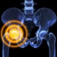 pathology of the upper femur, acetabulum, unpleasant, unbearable sensations caused by nearby soft tissue structures. It varies from mild to unbearable in terms of severity, dull, sharp, oppressive, painful, explosive, boring, etc. in nature. Can be. They often depend on the load, time of day and other factors. Causes of pain are determined using X-rays, CT, MRI, ultrasound, arthroscopy and other studies. Painkillers and rest of the limbs are recommended until diagnosis.
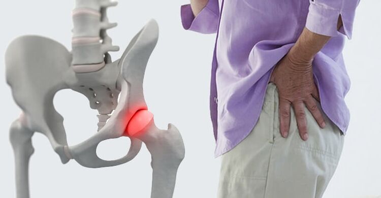
Causes of pain in the hip joint
Soft tissue injuries
The most common traumatic cause of pain is a contusion of the hip joint. Occurs when falling to the side or direct impact, manifests itself in moderate to severe pain, quickly becomes dull, gradually decreases and disappears within a few days, in severe cases - weeks. Support is maintained, movements are somewhat limited. Edema is found locally, bruising is possible.
Injuries to the hip joint are rare, usually the result of traffic accidents and sports injuries, accompanied by severe pain, and sometimes - a feeling of cracking (such as tearing tissue). The pain decreases slightly, then often increases again due to edema. Swelling from the joint extends to the groin and thigh.
The degree of dysfunction in the trauma of the ligaments depends on the severity of the injury (stretching, tearing, tearing) from the slightest limitation to the inability to stand. The pain increases with the deviation of the trunk, movements in the opposite direction of the damaged ligament.
Bone and joint injuries
Hip fractures usually occur in the elderly as a result of domestic or street trauma. A characteristic feature, especially in the presence of osteoporosis, is the absence of severe pain syndrome, mild edema. At rest, the pain is deep, dull, moderate, or insignificant, and the painful sensations increase sharply with movement. Support is sometimes maintained. A common symptom is the inability to lift a straightened leg in a prone position (symptom of a compressed heel).
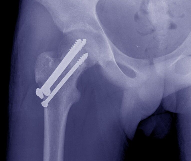
Transtrocanteric fractures are more commonly diagnosed in middle-aged and young people and develop as a result of high-energy trauma. Unlike cervical fractures, it is accompanied by unbearable sharp diffuse deep pain. Then the pain decreases, but remains very strong, difficult to tolerate. The joint is swollen, bruising is possible. Movement is very limited. Support is not possible.
Isolated fractures of the larger trochanter are rare; found in children and adolescents; It is caused by a fall, a direct impact or a sharp muscle contraction. The pain is sharp, very strong, mainly localized on the outer surface of the joint. Due to the increased pain, the patient avoids active movements.
Hip height occurs during falls, industrial and road traffic injuries, and manifests itself in unbearable sharp pains that do not decrease until they decrease. The joint is deformed, the leg is shortened, the knee joint is bent, and it turns outwards, less inwards (depending on the type of dislocation). Support and movement are not possible, summer resistance is determined when trying to move.
Acetabular fractures develop in isolation or are associated with hip dislocation. It is characterized by sharp explosive pain in the depths of the hip joint. Later, the pain subsides a little, but remains intense, impeding any movement. The leg is shortened and turned to the side. Support is not possible.
Degenerative processes
Pain with coxarthrosis in the early stages is a periodic, dull, indeterminate localization, which spreads to the hip, knee joint, sometimes at the end of the day or after a significant load. Light, fast-passing stiffness is possible at the beginning of the movement. Then the intensity of pain increases, painful sensations are noted not only during exercise, but also at rest. After severe stress, the patient begins to slow down. Movement is somewhat limited.
In severe coxarthrosis, the pain is deep, diffuse, persistent, painful, twisted. Worry both day and night. Decreased resistance to stress; while walking, patients lean on a cane. Movement is significantly limited, the affected leg is shortened, which increases the load on the joint, causing increased pain when walking and standing.
Chondromatosis of the hip joint resembles subacute arthritis. The pain is moderate, diffuse, transient, accompanied by tingling, limited mobility. When intraarticular bodies are damaged, blockages occur that are characterized by severe pain, impossibility, or significant restriction of movement. Once the joint mouse disorder has stopped, the listed symptoms disappear.
Trochanteritis is usually caused by osteoarthritis of the hip joint, accompanied by inflammatory-degenerative damage at the point of attachment of the tendons of the thigh muscles to the greater trochanter, manifested by pain in the area of the superficial lesion. position on the affected side. There is an increase in pain when trying to escape with hip resistance.
Bone malnutrition
Perthes' disease develops in children and adolescents, is characterized by partial necrosis of the femoral head, which is initially accompanied by a pale deep pain that is not intense, sometimes spreading to the knees and hips. After a few months, the pain intensifies sharply, becomes persistent, sharp, tiring. The joint swells, movement is limited, and lameness develops. Then the pain decreases, the rate of recovery of joint function changes.
Aseptic necrosis of the lower part of the femoral head is similar to Perthes' disease, but is found in adults, lasts less positively, and is bilateral in half of the cases. At first, the pain is intermittent. Then the pain syndrome intensifies and appears at night. At the peak of clinical manifestations, the pain is so intense that a person completely loses the ability to lean on his feet. Then the pain gradually subsides. Restriction of movement for about 2 years, resulting in arthrosis of the hip joint, contractures and shortening of the joint.
In 10-15-year-old boys, single bone cysts form in the proximal metaphysis of the thigh, which is accompanied by non-intense intermittent pain in the hip joint. There is no edema at all, long-term contractures often develop, especially in young children. The cause of treatment due to mild symptoms is a pathological fracture or increased restriction of movement.
Arthritis
Aseptic arthritis manifests itself as a wave-like pain in the joint that increases in the early hours of the morning. The severity of the pain varies from insignificant to severe, intense, persistent, significantly limiting physical activity. Hardness, swelling, redness and increased local temperature are noted. Palpation is painful.
Rheumatoid arthritis rarely involves the hip joints, the lesion is symmetrical. Periodic pain initially occurs against the background of changing seasons (autumn, spring), with a sharp change in weather conditions, hormonal changes after childbirth or during menopause. The pain is moderate or mild, diffuse, tingling or aching, increasing sharply on palpation. Recurrent synovitis is associated with edema, hyperemia, hyperthermia, increased mobility.
Infectious arthritis develops with the spread of hematogenous or lymphogenic infection, to a lesser extent - the penetration of the pathogen from nearby tissues into the joint. Acute onset with typically rapidly increasing pain. The pain is intense, twitching, tearing, exploding, restless, aggravated by movement, so the limb takes a forced position. Patients have fever, chills, sweating, severe weakness, edema, redness in the joints, and an increase in local temperature.
Without timely treatment, bacterial infectious arthritis can turn into panarthritis - a purulent inflammation of all tissues of the hip joint. It is characterized by a severe course with very sharp widespread throbbing pains, intense fever, severe weakness, pre-syncope, significant hyperemia and hyperthermia.
Other inflammatory diseases
Osteomyelitis of the upper thigh can be hematogenous, post-traumatic or postoperative. Hematogenous osteomyelitis manifests itself with very sharp bursting, twitching, tearing, or dull pain, clearly localized as the patient avoids the slightest muscle movements. There is hyperthermia, severe intoxication.
Post-traumatic and postoperative osteomyelitis occur with similar but less pronounced symptoms. Typically, the appearance of a more gradual onset, purulent discharge on the background of an open fracture or postoperative wound. Pain in the hip joint increases in 1-2 weeks in parallel with the development of local inflammatory symptoms.
Synovitis develops against the background of injuries, other diseases of the hip joint, there is less manifestation of allergies. In acute synovitis, the pain is usually small, dull, exploding, and gradually increases due to an increase in the amount of articular fluid. The joint is swollen, palpation is slightly painful, there is a sign of fluctuations. Chronic synovitis is asymptomatic, accompanied by mild excruciating pain.
Pain with intermediate hydroarthritis is also insignificant, accompanied by restlessness, limited mobility, and disappears within 3-5 days after reverse resorption of effusion. They are updated at individual intervals for each patient, causing recurrent fluid accumulation in the joint.
Specific infections
Tuberculosis of the hip joint is a common form of osteoarticular tuberculosis, manifested by general weakness, fatigue, subfebrile condition. Then there are weak pulling or aching pains in the muscles, temporary pain in the joints when walking. The patient begins to spare the limb. As the pain progresses, it spreads moderately, spreads to the knee, and is accompanied by swelling, redness, and synovitis. Develops a protective contracture.
Joint pain, including hips, may appear with brucellosis. Painful sensations associated with acute and subacute withdrawal, torsion, intermittent fever, lymphadenopathy, skin rash. Pain syndrome in a chronic course is similar to the formation of deformities over time in aseptic arthritis.
Congenital anomalies
Manifestations of hip dysplasia are determined by the degree of mismatch of the femoral head and acetabulum. With a complete congenital dislocation, the pain is accompanied by lameness immediately after the child begins to walk. With moderate subluxation, painful sensations occur at the age of 5-6 years, which is directly related to the load on the foot.
With a mild subluxation, the pathology is asymptomatic for a long time, the pain syndrome manifests itself with the development of dysplastic coxarthrosis at the age of 25-30 years. Symptoms of such osteoarthritis include rapid onset of pain, early onset of pain at rest and at night, and progressive restriction of movement. All forms of dysplasia are accompanied by asymmetry of skin wrinkles, "click" symptoms and limited mobility. In the case of dislocation, shortening of the joint is noted.
Neoplasms
A typical asymptomatic course for benign neoplasms. The pain is small, intermittent, and often does not last for years. Tumor growth is accompanied by a slow increase in pain syndrome, recurrent synovitis. Osteomas, osteoid osteomas, osteoblastomas, chondromas are more common in the hip joint.
Malignant neoplasms (osteosarcomas, chondrosarcomas) are characterized by the rapid development of pain syndrome and other pathological manifestations. Initially, the pain is small, short-lived, without a specific localization, and sometimes more severe at night. Then they become sharp, durable, cutting, encircling, spreading to the entire joint. The affected area swells and deforms. Weight loss, weakness, subfebrile condition are noted. Painful, unbearable pain with advanced neoplasms can be eliminated only with drugs.
Other reasons
Pain in the hip joint sometimes occurs with lumbosacral plexitis and sciatic nerve neuropathy, however, they usually occupy an insignificant position in the clinical picture of the disease, with severe pain in the back and thighs in the background compared to limb weakness. and sensitivity disorders.
Pain syndrome of this localization is often found in osteochondrosis and disc herniation. Pain can be detected with spondylitis, deformed spondyloarthritis and curvature of the spine. The pains are dull, periodic, painful, often intensifying during the exacerbation of the underlying disease. Their appearance may be due to a constant overload of the joint or the development of coxarthrosis.
Sometimes joint pain is caused by mental illness or depressive illness. Diabetes mellitus is often accompanied by entesopathies, capsulitis and other lesions of the periarticular soft tissues. Possible drug arthropathy when taking certain medications.
Diagnostics
In case of injury, diagnostic measures are performed by traumatologists. Degenerative and inflammatory diseases are managed by orthopedists and rheumatologists. The participation of surgeons in purulent processes is important. The examination includes the collection of complaints, study of medical history, physical examination, additional research. The following methods can be used, taking into account the characteristics of the pathological process:
- Radiography.It is a basic technique for most joint diseases. Detects fractures, dislocations, changes in the contour of the acetabulum and femoral head, peripheral and intraosseous defects, bone growth, narrowing of the joint space.
- Ultrasound.It is most informative when studying soft tissues. Detects signs of inflammatory and degenerative processes, areas of calcification. Used to diagnose eruptive, articular mice.
- MRI and CT.In order to clarify the nature, prevalence and location of the pathological focus, clarification methods are used when there is ambiguous information from the main research. Can be done with contrast.
- Puncture of the joint.Diagnostic or therapeutic and diagnostic. It allows to eliminate the effusion, to study the composition of the intra-articular fluid, to determine the causative agent of infection using laboratory tests.
- Arthroscopy.During the visual examination of the joint, the doctor assesses the condition of the bone and soft tissue structures, if necessary, takes a biopsy sample for further histological examination and performs therapeutic measures.
- Laboratory tests.It is prescribed to determine the signs of inflammation and markers of rheumatic diseases, to assess the general condition of the body, the activity of various organs in severe infectious or systemic pathologies.
Treatment
Please help before making a diagnosis
In severe injuries, it is necessary to straighten the joint by applying a tire from the foot to the armpit. In the case of minor traumatic injuries, it is sufficient to provide the foot with rest. Cold is applied to the affected area. An analgesic is given for severe pain. You can not do active movements with the muscles, load the leg. It is strictly forbidden to try to prevent the bones from moving and moving.
The tactics of non-traumatic illness are determined by symptoms. With small manifestations, the use of local remedies with analgesic and anti-inflammatory effect is allowed to ensure the relaxation of the joint. In case of fever, weakness, severe pain, edema and rapid growth of hyperemia, it is recommended to seek specialized help immediately.
Conservative therapy
Speeches are an indication of an immediate decline. Fractures are usually treated with skeletal traction, then the patient is operated on or the limb is fixed with a cast after the callus symptoms appear. Elderly patients with a hip fracture are allowed immobilization with a derotating pull, which prevents rotational movements in the joint.
The remaining patients are advised to relax the hip joint. Depending on the indications, it is recommended to use orthoses or additional devices (crutches, canes). Assign massage, physiotherapy exercises, physiotherapy procedures:
- laser therapy;
- magnetotherapy;
- UHF;
- ultrasound;
- electrophoresis with drugs;
- UHT.
It is possible to use NSAIDs, chondroprotectors, antibacterial drugs. Local agents are widely used. Indications for joint holes, intra- and periarticular occlusions with hormones, intra-articular injections of chondroprotectors, synovial fluid substitutes.
Surgery
Operations on the hip joint are performed with the help of open access and arthroscopic devices. Depending on the type of pathology, the following can be done:
- Traumatic injuries:open reduction of hip dislocation, reconstruction of the acetabulum, cervical osteosynthesis, trochanteric fractures.
- Degenerative processes:arthrotomy, arthroscopy, removal of free intramuscular organs.
- Tumors:removal of neoplasia, bone resection, rupture of the hip joint, amputation of the abdominal cavity, resection of the abdominal cavity.
In case of contractures, ankylosis, scarring in periarticular tissues, rehabilitation, arthroplasty and arthrodesis are performed. Endoprosthetics are an effective way to restore limb function in diseases of various origins accompanied by restriction of movement or destruction of the joint.